Case Study: UC Berkeley CellScope
Mobile Medical Microscopy
Key Metrics
2 Devices
A retinal camera and a TB microscope, engineered end-to-end
5 Continents
CellScope platform piloted in global-health settings
100°
Widefield retinal coverage, up from 20–40° per capture
4 Papers
Peer-reviewed clinical publications
Origins in Academic Innovation
In 2009, as smartphones were beginning to feature high-resolution cameras for the first time, Professor Dan Fletcher's Bioengineering lab at UC Berkeley was among the first to systematically apply these consumer optics to medical diagnostics.
"Cell phones are very widespread in the developing world. 90% of the global population lives within mobile internet coverage, and many of those places never had landline communication or reliable electricity."
— Dr. David Breslauer
CellScope, first published by David Breslauer in 2009, rested on a deceptively simple insight: the image sensors being engineered into phones so people could take better photos were, with the right optical attachment, capable of capturing diagnostic-quality microscopy. With a small, low-cost clip-on lens and purpose-built software, a clinician anywhere in the world could perform laboratory-grade imaging in the field.
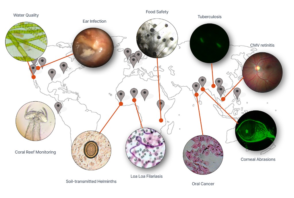
Over the following years, the Fletcher Lab built a wide range of prototype mobile microscopes, each exploring a different clinical application: tuberculosis, retinal disease, bloodborne parasites, ear infections, oral cancer screening, water-quality monitoring, and education. The unifying thread was a vision of democratizing diagnostics—leveraging ubiquitous consumer hardware to push clinical capability out to the places where it was needed most.
What made CellScope more than a series of demos was its commitment to academic translation through pilot studies: taking each prototype into the field, into the hands of real clinicians, and measuring it against established gold standards. That is a fundamentally different discipline from publishing a proof of concept—and it is the discipline that separates a promising idea from a deployable product.
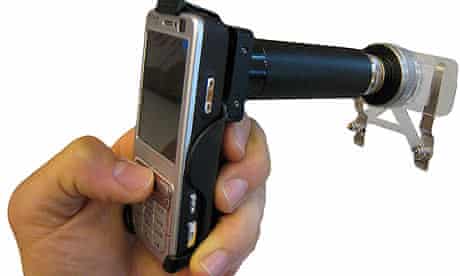
Mosaic's founding team did this translational work firsthand. As a research engineer in the Fletcher Lab, Mosaic founder Dr. Frankie Myers designed and built the devices, developed all of the firmware and iOS software, worked with in-country partners and physicians to pilot and evaluate them against established gold standards, and connected the lab with manufacturing vendors who could scale them. That end-to-end path—from bench prototype to field-validated, manufacturable device—is exactly the work Mosaic was built to accelerate.
The Leapfrog Effect
In technology, a "leapfrog" occurs when a region bypasses an older generation of infrastructure and jumps straight to the next—rural communities that never had landline phones went directly to mobile. CellScope applied the same logic to diagnostics: instead of building out expensive laboratory infrastructure, bring the lab to people's phones. AI now represents the next, and perhaps more dramatic, leapfrog: pushing clinical intelligence to the point of care worldwide.
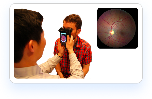
Mosaic was involved in two applications of mobile medical microscopy from the Fletcher Lab: Ocular CellScope —a smartphone-based retinal camera, and Tuberculosis CellScope —an AI-assisted automated slide-scanning microscope. Together they demonstrate how diagnostic-quality imaging can be delivered almost anywhere using mobile devices and intelligent software.
Ocular CellScope (Retinal Imaging)
Smartphone-based retinal imaging for diabetic retinopathy screening.
Tuberculosis CellScope (Automated Slide-Scanning Microscope)
AI-assisted mobile microscopy for tuberculosis detection.
Global Pilots
Project One
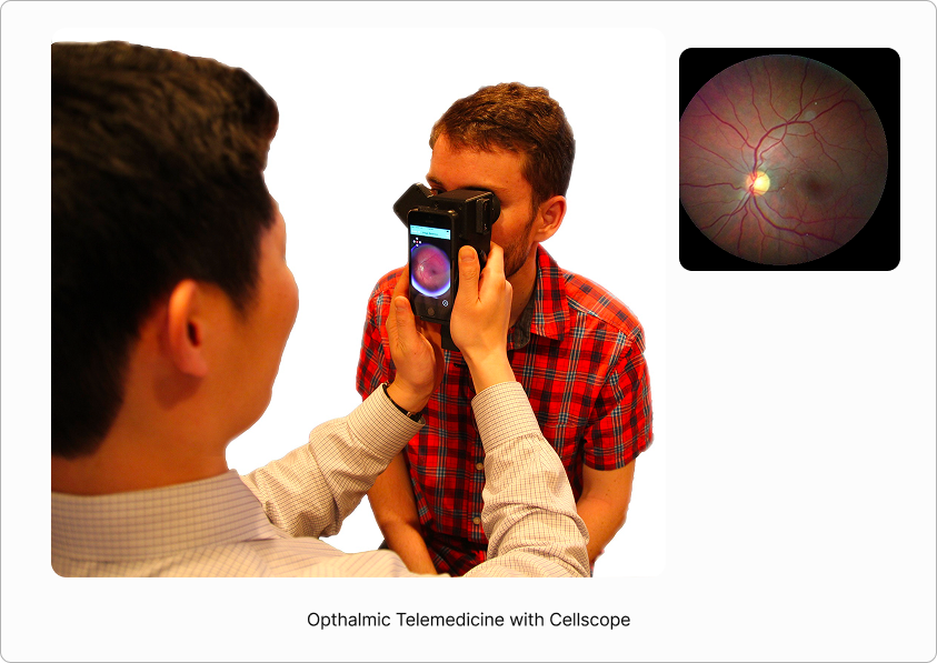
Ocular CellScope: Retinal Imaging
A handheld, smartphone-powered retinal imaging system for accessible eye diagnostics
The Access Crisis in Diabetic Retinopathy
Diabetic retinopathy is the leading cause of vision loss in working-age adults, yet it is highly treatable when caught early. The tragedy is one of access: nearly half of diabetic adults do not receive recommended annual screening, and in vulnerable populations with limited access to specialty care, screening rates fall to just 10–20% per year.
643M
Projected adults living with diabetes by 2030 (IDF Diabetes Atlas)
~50%
Of diabetic adults miss recommended annual eye screening
10–20%
Annual screening rate in low-access populations
Technical Challenges in Smartphone-Based Imaging
Early smartphone retinal cameras showed promise but faced critical limitations that prevented clinical adoption. Ocular CellScope was designed from the ground up to systematically address each one.
Limited Field of View
Problem: Gold-standard DR screening requires seven standard fields to cover the retina. Single smartphone images typically capture only 20–40°.
Why it matters: incomplete coverage misses peripheral lesions and reduces screening sensitivity.
Patient Discomfort
Problem: Sustained bright white illumination causes pupil constriction, glare, and patient movement.
Why it matters: motion artifacts and poor localization lead to repeat exams and lower image quality.
Operator Skill Required
Problem: Surveying wide retinal regions demands experience to direct gaze and capture overlapping fields systematically.
Why it matters: inconsistent coverage limits scalability in resource-limited settings.
Workflow Complexity
Problem: Image acquisition, transfer, stitching, and review often span multiple devices and steps.
Why it matters: this delays feedback, increases cost, and prevents point-of-care decision-making.
Portability
Problem: Tabletop fundus cameras are large, mains-powered, and require a seated, cooperative patient—impractical at the bedside, in the field, or with children and critically ill patients.
Why it matters: portability decides whether screening reaches the people who need it most, or stays confined to specialty clinics.
The Automation-First Strategy
"We weren't just miniaturizing a fundus camera. We were reimagining the entire workflow around what automation could enable."
— Frankie Myers, PhD
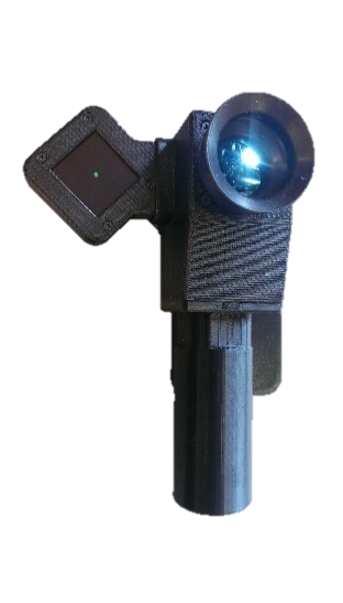
Automated Fixation Guidance
A software-controlled external display presents a moving target that guides the patient's gaze through a standardized sequence, enabling consistent widefield coverage without operator expertise.
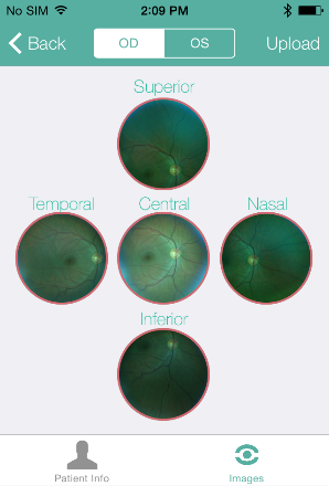
On-Phone Montage Stitching
Custom OpenCV/SURF algorithms run entirely on-device to align and stitch multiple captures into a single diagnostic-quality widefield image.
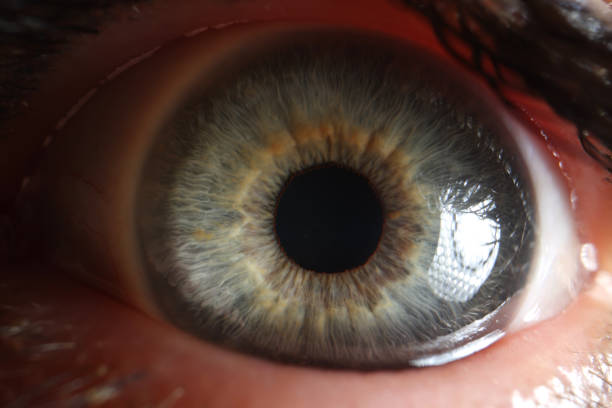
Dual-Wavelength Illumination
Far-red (655nm) preview LEDs keep patients comfortable during alignment; standard illumination is used only for capture, reducing discomfort and motion artifacts.
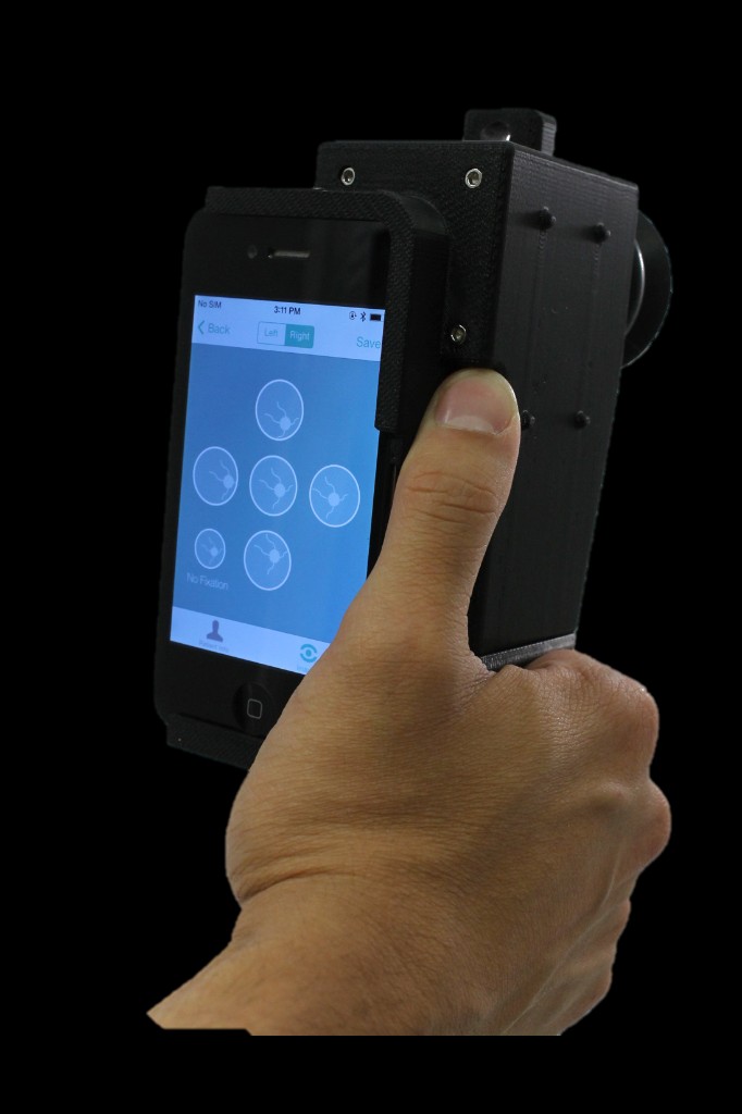
One-Handed Ergonomic Design
A symmetric housing with a magnetic fixation-display attachment allows one-handed operation and stable imaging across varied clinical settings.
Hardware Architecture
Every component was purpose-designed for portability, patient comfort, and clinical-grade image quality.
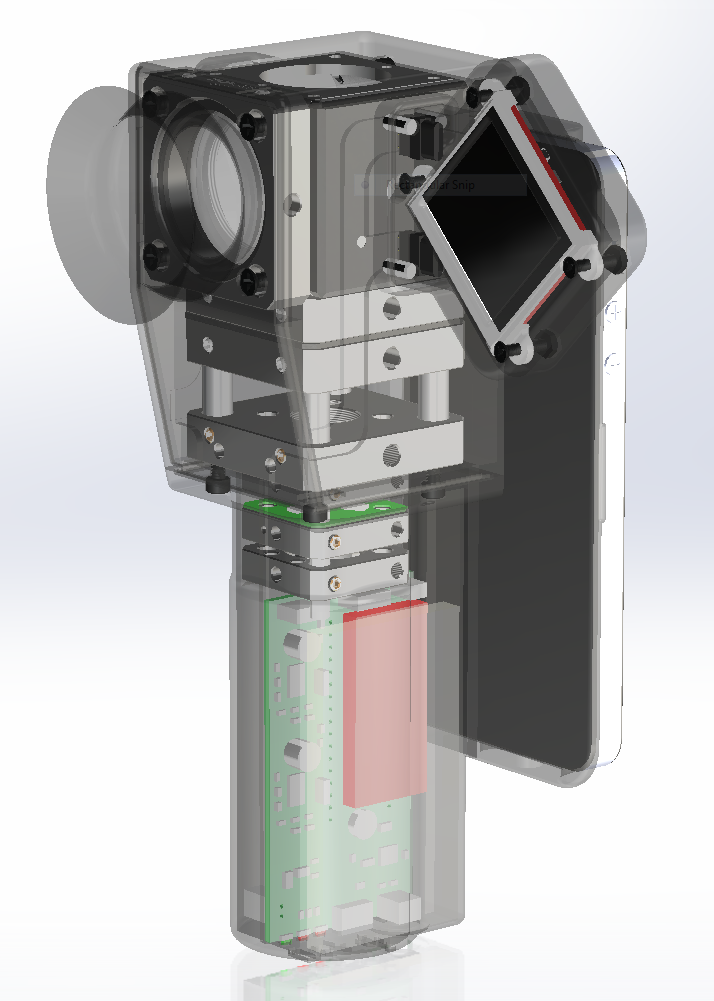
Optical system and electronics
- Polarizing wire-grid beam splitter minimizes corneal reflections.
- Annular illumination (4.8–9.6mm) at the corneal surface.
- 50° individual field of view per capture.
- Rechargeable Li-Po battery (7+ days typical use).
- Magnetic OLED fixation display with spring-loaded contacts.
Software Intelligence: The Breakthrough
Ocular CellScope was the first smartphone retinal imaging system to perform automated montage stitching entirely on-device—eliminating the need for external computers and enabling true point-of-care deployment.
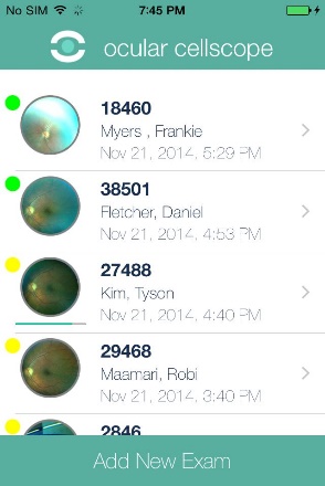
Patient Data Entry
Enter demographics and exam type. Data stays on-device until you choose to upload.
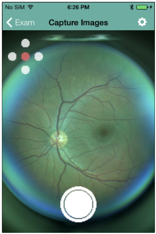
Real-Time Preview & Adjustment
Swipe to adjust focus, zoom, and exposure until the retina is clear. Far-red preview keeps patients comfortable.
Automated 5-Field Sequence
Fixation guides central, superior, inferior, temporal, and nasal fields—you capture at each stop.
On-Phone Image Stitching
OpenCV/SURF aligns the fields into one 100° widefield montage, entirely on the phone.
Review & Cloud Upload
Pinch, zoom, and swipe to review. Optional secure upload to an EHR or reading center.
Clinical Validation
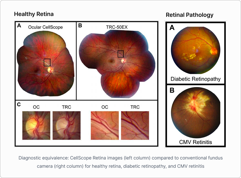
Ocular CellScope's real innovation emerged in the settings where traditional cameras simply can't go: emergency rooms, hospital beds, wheelchair-bound patients, and pediatric exams. Independent grading found its images diagnostically equivalent to conventional fundus cameras across healthy retinas, diabetic retinopathy, and CMV retinitis—and a follow-on study showed automated grading on par with expert human graders.
Project Two
Tuberculosis CellScope: Automated Slide-Scanning Microscope
An integrated platform for AI-assisted fluorescence TB screening in resource-limited health systems
~2B
People living with latent TB infection worldwide (WHO)
10.8M
People who fell ill with TB in 2023 (WHO Global TB Report)
~1.25M
TB deaths in 2023 (WHO Global TB Report)
Up to 50%
Of pulmonary TB cases missed by conventional smear microscopy
WHO ASSURED Criteria
Millions face barriers to early TB detection. Tuberculosis CellScope was designed against the WHO ASSURED framework for diagnostics built for last-mile care.
Affordable
Leverages consumer smartphone/tablet optics and global supply chains—a leapfrog effect for accessibility.
Sensitive
Matches the diagnostic accuracy of trained human microscopists in peer-reviewed clinical evaluation.
Specific
Detects TB bacilli with precision, minimizing false positives in community screening.
User-friendly
Operable by technicians with minimal training, with a localized interface for ease of use.
Rapid & Robust
Returns diagnostic results in roughly 20 minutes—actionable care at the point of need.
Equipment-free
Portable and battery-powered, bringing lab-grade capability to sites without fixed infrastructure.
Deliverable
Designed for last-mile delivery and scalable to remote and rural health centers.
The Platform
Tuberculosis CellScope wasn't just a microscope. It was an integrated diagnostic platform—hardware, software, and AI working together to reimagine the entire screening workflow.
Hardware Design
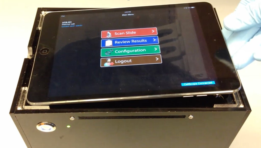
iPad-based system
Camera for imaging, display for visual confirmation, mobile connectivity for QC and telemedicine.
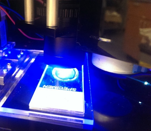
Fluorescence optics
Blue-light excitation for auramine-stained fluorescence microscopy.
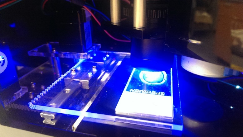
Automated slide scanning
Motorized stage scans the slide and captures fields without operator intervention.
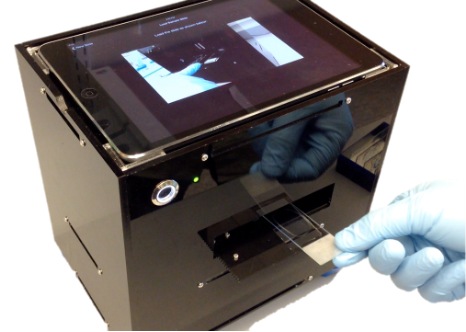
Rugged, compact enclosure
Battery-powered and field-deployable.
AI / Software Visualization
Scroll to move through the on-device analysis sequence.
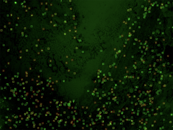
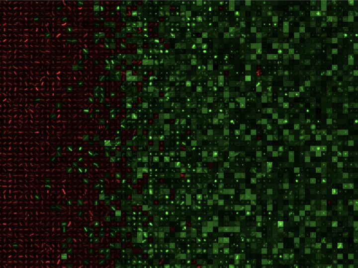
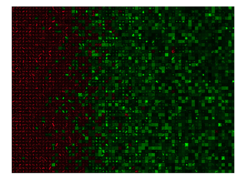
Raw capture — single-cell resolution fluorescence microscopy, no annotations.
AI classification — candidate bacilli marked with colored bounding boxes.
Scored output — detections sorted and ranked for rapid diagnostic review.
Reflections and Legacy
Lessons Learned
The CellScope years were formative. The platform proved that smartphone-based microscopy could meet clinical standards in the field—and it also revealed how much lies beyond the hardware when turning research into impact. Those lessons are foundational to how Mosaic works today.
Proof of Concept Is Not a Product
CellScope demonstrated across five continents that smartphone-attached microscopy could perform at clinical standards. But demonstrating something and shipping it are entirely different disciplines—requiring manufacturing scale-up, regulatory strategy, distribution networks, and pricing models that academic labs are not built to navigate.
Partnerships Are the Platform
The power of CellScope was never just the optics. It was the constellation of clinical partners, Ministries of Health, public-health departments, and international NGOs that the Fletcher Lab convened. Platform technologies derive their value from the relationships built around them, and sustaining those partnerships is a strategic capability in itself.
The Translational Pipeline Must Exist
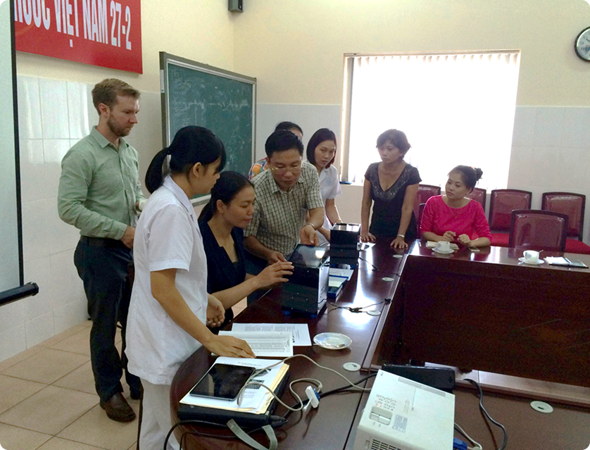
The Fletcher Lab could keep innovating—piloting in Vietnam, publishing, presenting, talking to clinicians worldwide. What was missing was a downstream translational pipeline: an organization that could take a validated prototype and carry it through commercial development. Without that handoff, impact stays perpetually out of reach. Mosaic exists to be that handoff.
Technology Alone Does Not Define the Right Solution
The TB CellScope was a remarkable instrument—and also a complex electro-mechanical system with many parts and significant cost of goods. In parallel, molecular diagnostics were emerging with higher sensitivity, lower complexity, and the ability to detect drug resistance. The lesson: always evaluate technology choices against the full landscape of solutions. Complexity is a liability at the last mile.
Research papers
Publications
Mobile Phone Based Clinical Microscopy for Global Health Applications
D.N. Breslauer, R.N. Maamari, N.A. Switz, et al. — PLoS ONE 4(7): e6320 (2009)
The original CellScope paper, demonstrating brightfield and fluorescence microscopy on a mobile phone, with proof-of-concept imaging of malaria parasites, sickle red blood cells, and tuberculosis bacilli—plus automated bacillus counting.
A Smartphone-Based Tool for Rapid, Portable, and Automated Wide-Field Retinal Imaging
T.N. Kim, F. Myers, C. Reber, et al. — Transl Vis Sci Technol 7(5): 21 (2018)
Foundational Ocular CellScope paper. Introduces a smartphone-based fundus camera with a detachable, programmable fixation display and automated stitching of multiple captures into wide-field retinal composites.
Comparison of automated and expert human grading of diabetic retinopathy using smartphone-based retinal photography
T.N. Kim, F. Myers, et al. — Eye 35, 334–342 (2021)
Demonstrates that automated grading of smartphone-captured retinal images performs on par with expert human graders for diabetic retinopathy—closing the loop from portable capture to point-of-care interpretation.
Evaluation of mobile digital light-emitting diode fluorescence microscopy in Hanoi, Vietnam
L.H. Chaisson, C. Reber, N. Switz, et al. — Int J Tuberc Lung Dis 19(9): 1068–1072 (2015)
Field evaluation of the CellScope TB instrument in Hanoi, Vietnam, assessing diagnostic performance when operated by non-specialist personnel, with automated image upload over mobile networks for centralized review.
Acknowledgments
Dan Fletcher
Principal Investigator · UC Berkeley
Clay Reber
Research Scientist · UC Berkeley
David Breslauer
UC Berkeley / UCSF Bioengineering
Robi Maamari
UCSF Ophthalmology
Tyson Kim
UC Berkeley Bioengineering
Neil Switz
UC Berkeley Bioengineering
Payam Nahid
UCSF Curry TB Center
Adithya Cattamanchi
UCSF / SF General Hospital
Nguyen Van Nhung
National Lung Hospital · Hanoi
Ha Phan
CPAS · Hanoi, Vietnam
Jeannette Chang
AI Algorithm · UC Berkeley
Jitendra Malik
AI Algorithm · UC Berkeley
"CellScope taught us that the hard part of global-health diagnostics isn't the demo—it's everything after: the pilots, the partnerships, the manufacturing, and the path to scale."
Mosaic Design Labs — Dr. Frankie Myers, PhD
Have a lab technology ready to translate?
Let's turn a validated prototype into a manufacturable, field-ready product.
Explore Projects